Immunofluorescence with Vector Laboratories
A summary of Immunofluorescence from Vector Laboratories

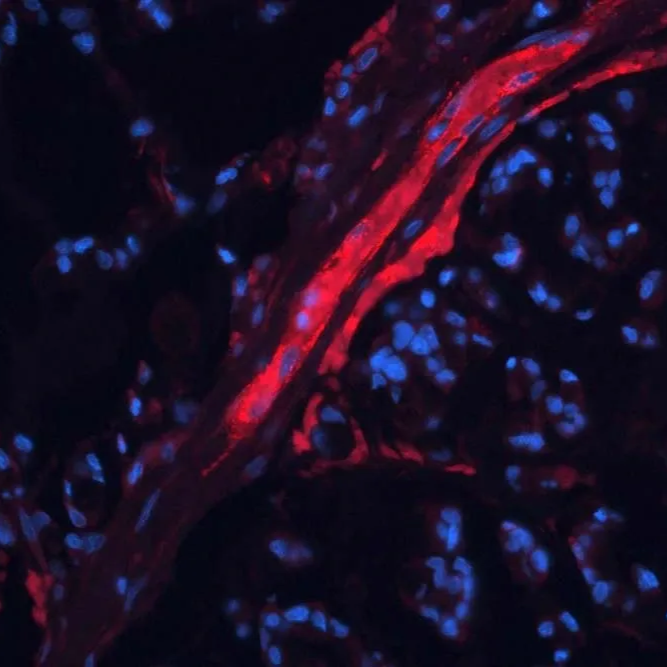
What is Immunofluorescence?
Immunofluorescence is a technique that uses antibodies to detect proteins of interest in tissue sections with a fluorescence output. It needs a fluorescent or confocal microscope to enable visualization. It’s often used as an alternative to immunohistochemistry for several reasons.
- Does not rely on enzymes, which can avoid issues with endogenous enzymes.
- The fluorophoresare smaller than depositing substrates used in IHC and allow for better resolution without the issue of steric hinderance.
Vector Laboratories has been developing and manufacturing reagents for immunofluorescence for nearly 50 years, and their products are known for their reliability and reproducibility.
The primary antibody is usually detected with secondary antibodies either directly conjugated with a fluorophore or to biotin for further detection with a variety of avidin or streptavidin labelled with fluorophores.
Here is an outline of the IF method, which shows possible steps and reagent choices:

Here is an outline of the IF method, which shows possible steps and reagent choices
How To Get The Best Results From Your Immunofluorescence
There are a lot of variables that can affect your IF staining, and these can be considered in the design stages to reduce the amount of optimization needed.
Tissues
Tissues used are most commonly FFPE or frozen sections. Not all primary antibodies will work on both tissue types, and some antigens can be destroyed by fixation methods, so where possible start by matching your fixation to your primary antibody’s needs.
Further information on Sample preparation can be found in this guide.
Formalin fixation can cause tissues to autofluoresce. The issue can be mostly avoided by using different fixatives, or the TrueView Autofluorescence quenching kit can be used at the end of the protocol.
Primary antibodies
As mentioned in "Tissues", ensure that your primary antibody is known to recognise your antigen in your type of fixed tissue.
You should also ideally choose a primary antibody of a different species to your tissue if possible. If you can’t, you may need to consider a conjugated primary antibody, or a kit designed to eliminate cross reactivity. Our Technical Team are happy to discuss your case.
Antigen Unmasking
Some antigens need unmasking to enable the primary antibody to recognise them. Techniques such as enzyme digestion or Antigen Unmasking/ Heat Induced Epitope Retrieval (HIER) are often used. This step is usually dictated by the primary antibody to be used and is most commonly required on FFPE samples.
Secondary antibodies
Secondary antibodies offer a flexible way to detect a primary antibody, and also help to increase the sensitivity of the method, although they can also contribute to background staining, especially if the primary antibody is closely related to the tissue species – e.g. mouse and rat. The secondary antibody should be chosen to detect the primary antibody, and antibodies adsorbed against the tissue species are recommended if there’s a cross-reactivity issue. The secondary antibody needs to either have a hapten, such as biotin or the fluorophore conjugated to it. Tyramide fluorophores are becoming a popular method for multiple labelling, these require a secondary antibody with HRP. Kits such as ImmPRESS® or VECTASTAIN® can be used in conjunction with tyramide for an exceptionally sensitive method.
Choosing Fluorophores
There are a wide range of fluorophores now available.
- Choose fluorophores need to be resistant to photobleaching for imaging tissues and cells, as signals can quickly fade during visualization.
- Check the excitation and emission spectra are compatible with the fluorescence microscope being used to be able to read results.
- Compare fluorophores with different excitation and emission spectra for multiple labelling/ multiplexing and choose fluorophores with little/no overlap. Computer programmes can be used to unmix some fluorophores, but it can be easier to avoid this as a necessity if possible.
2B Fluorophore Table - Check fluorophore brightness and wavelength penetration. Brighter fluorophores are more sensitive, however there can also be a benefit to using a longer wavelength fluorophore in thicker sections and Z-Stacking, as the longer wavelengths can sometimes penetrate and escape from tissues better than the brighter, but shorter wavelength alternatives.
Mounting
To help preserve fluorophores, an ani-fade mounting media is recommended. There are a wide variety of Vectashield Mounting Media that offer setting or non-setting formulations, as well as the option of different counterstains, such as DAPI, PI or TRITC-Phalloidin that can help highlight cellular architecture.
Further information on mounting sections for IF can be found in this resource.
Frequently asked questions
Immunofluorescence Technical Resources
Immunofluorescence Resource Guide
This guide will help you:
- Learn about the IF workflow and how to optimise each step.
- Quickly identify and choose the most appropriate fluorescent secondary conjugates.
- Select the best anti-fade medium to preserve your fluorescence signal for imaging and archiving
TrueVIEW Autofluorescence Quencher Brochure
For those wanting additional details on the simple protocol and mode of action of the TrueVIEW Autofluorescence Quenching Kit, this brochure contains schematics, comparison data with other types of quenchers, and customer testimonials.
Immunofluorescence Mounting Guide
This IF mounting guide provides information to help researchers choose the best mounting medium for their project and offers tips to help reduce photobleaching during the image visualization process.
Find out more about this hardening anti-fade mounting media that's great across the whole spectrum.
Vector's latest anti-fade mounting media that's great across the whole spectrum, but in a non-setting formulation.
FFPE or Frozen? How to prepare your samples for staining.
Talk to an IF specialist
If you have any further questions please contact us to arrange a call with our in house IF specialist.