Skip to content

Neuroscience research tools for imaging, neurodegeneration and signalling

With around 86 billion neurons and almost a quadrillion synaptic connections, the human brain is extraordinarily complex (1). Neuroscience aims to unravel this complexity, from visualising brain structure and neural cells to understanding neuronal signalling and the mechanisms underlying neurodegenerative disease.

Investigating these processes requires diverse research tools, from imaging reagents and antibodies to disease models and pathway modulators. In this blog, we explore the neuroscience tools available through 2BScientific from our trusted specialist suppliers, supporting neural imaging, neurodegeneration modelling and research into altered signalling in disease.
Neuron-stock-image

Visualising proteins and cell types in neuroscience

The nervous system is built from a dazzling variety of specialised cells, each defined by the proteins it expresses. Understanding where these cells and proteins are located and how this organisation shifts in health and disease, is fundamental to neuroscience.

Immunohistochemistry (IHC) and fluorescence microscopy give researchers a method to see this organisation directly. By tagging specific proteins within neural tissue, researchers can identify neurons, astrocytes and microglia, distinguish progenitor from dying cells and map neuropeptide and neurotransmitter systems. Significantly, these techniques make it possible to compare how protein expression and localisation change between healthy and diseased tissue.

This type of protein visualisation is particularly valuable in neurodegeneration research. For example, pathological hallmarks such as amyloid beta and tau in Alzheimer’s disease, and alpha synuclein in Parkinson’s disease, can be visualised to investigate disease associated changes.

Importantly, the choice of antibody and detection method can influence how clearly these targets are visualised. HistoSure antibodies from SYSY Antibodies are developed for formalin-fixed, paraffin-embedded tissue, supporting protein detection in preserved samples.

For higher-precision fluorescence imaging, FluoTags from NanoTag Biotechnologies offer a much smaller alternative to conventional IgG antibodies. Their compact size improves tissue penetration and epitope access, while positioning fluorophores closer to the target for more precise localisation.
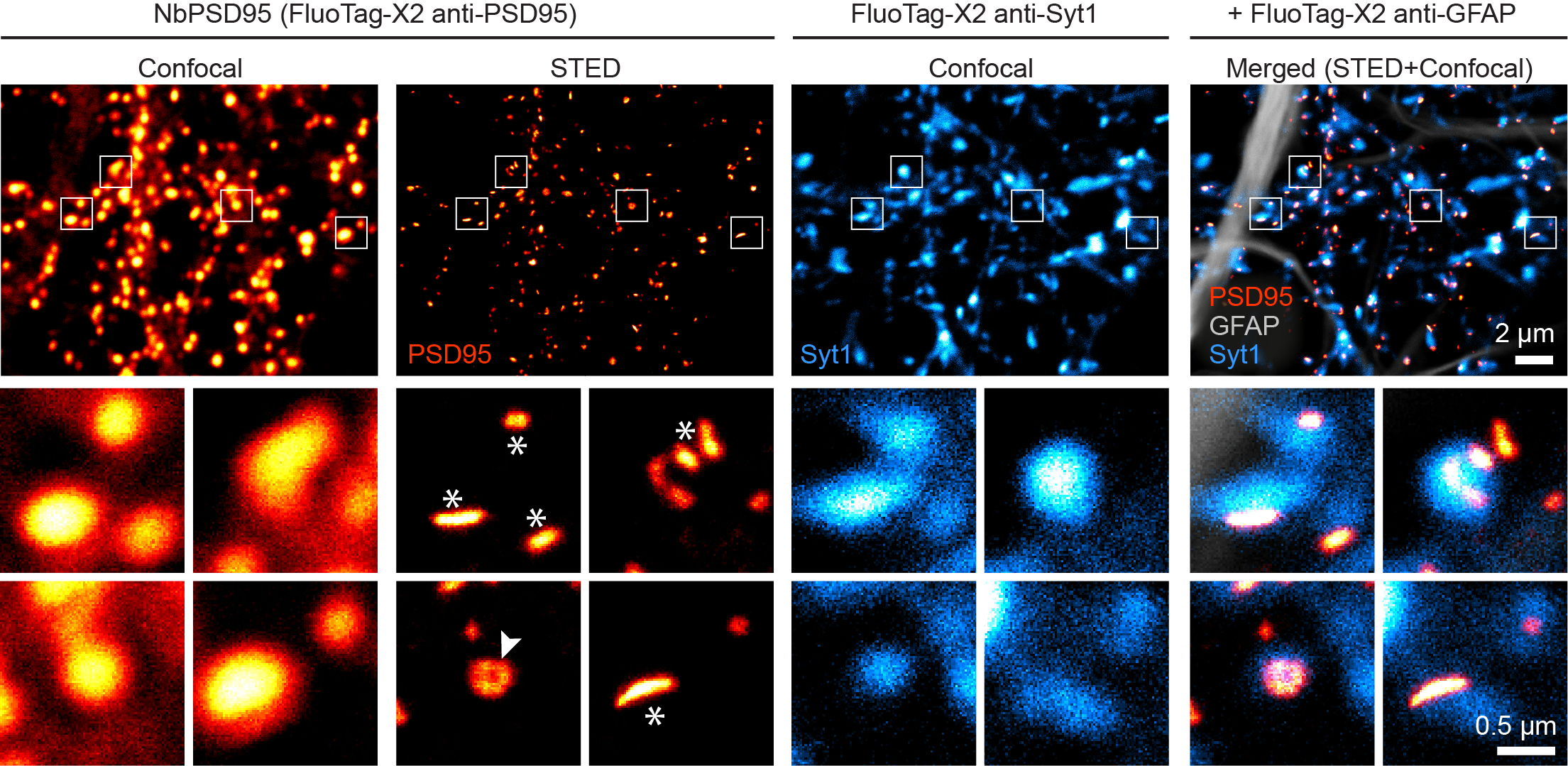
Fig4

Figure 1 - FluoTag® imaging of hippocampal neurons highlights synaptic and glial proteins, with STED microscopy revealing finer synaptic detail than conventional confocal imaging.

Modelling neurodegeneration and protein aggregation

Many neurodegenerative diseases are characterised by progressive neuronal loss alongside abnormal protein accumulation. In Alzheimer’s disease, extracellular amyloid beta plaques and intracellular tau tangles are major pathological hallmarks (2). Conversely, Parkinson’s disease is associated with the loss of dopaminergic neurons and the intracellular accumulation of alpha synuclein within Lewy bodies (3).

To understand how these pathologies develop and assess potential interventions, researchers need experimental models that reproduce key features of disease. Pre-formed fibrils (PFFs) provide one way to do this. Once introduced into a cellular or animal model, PFFs act as “seeds” encouraging the cell’s own corresponding proteins to misfold and form abnormal aggregates (4,5). This allows researchers to reproduce and follow disease-associated protein aggregation under controlled experimental conditions.

StressMarq Biosciences offers PFFs for modelling several forms of neurodegenerative protein aggregation:

  • Alpha synuclein PFFs can induce disease-like alpha synuclein pathology relevant to Parkinson’s disease
  • Tau PFFs can seed tau aggregation for Alzheimer’s disease and other tauopathies
  • SOD1 PFFs support studies of SOD1 aggregation associated with amyotrophic lateral sclerosis (ALS).

These models enable researchers to investigate how aggregation develops, its effects on neural cells and how potential interventions influence the process.

Beyond protein aggregation, neurodegeneration also involves changes in cellular stress responses and neurotransmitter signalling. Small molecules can help researchers probe these pathways. Focus Biomolecules offers tools such as ISRIB, an integrated stress response inhibitor used in amyloid beta and tau-related research and apomorphine, a dopamine receptor agonist relevant to Parkinson’s disease models.

StressMarq Biosciences

Established in 2007, StressMarq Biosciences Inc. is a supplier of life science products that operates out of Victoria, Canada with a small, but dedicated group of scientists. Headed by the CEO and President Dr. Ariel Louwrier, StressMarq provides the research community with high-quality reagents backed with rigorous quality control data, expert scientific support, and fast international delivery.

Investigating neuroinflammation, neuronal stress and signalling

Although abnormal protein aggregates are well-known hallmarks of Alzheimer’s and Parkinson’s disease, neuronal damage can emerge from a network of interacting processes involving inflammation, disrupted signalling, oxidative stress and cell death (6).

Neuroinflammation is driven largely by inflammatory responses from glial cells, while excessive glutamate signalling can cause excitotoxic neuronal damage (7). Notably, oxidative stress can also contribute to ferroptosis, an iron dependent form of regulated cell death. Rather than occurring independently, these processes can interact and amplify neuronal dysfunction as disease progresses.

Small molecules allow researchers to selectively perturb these pathways and examine how neurons and glial cells respond. Focus Biomolecules provides compounds spanning several of these mechanisms. Ibudilast can support studies of neuroinflammation and glial cell activation, kainic acid is widely used to induce neuronal excitation and model excitotoxicity, while UAMC-4821 enables researchers to investigate ferroptosis and its contribution to neuronal dysfunction.

2BScientific also supplies a broad range of antibodies against relevant neuroscience targets and biomarkers, allowing researchers to examine proteins, cell populations and signalling responses alongside these experimental models.

Together, imaging reagents, disease models, small molecules and antibodies provide complementary ways to investigate the complex biology underlying neurological disease.

Focus Biomolecules

Focus Biomolecules was founded by former members of globally recognised BIOMOL. From their industry experience they set out to refine the practices of small molecule based life sciences companies and eliminate the challenges too often found by customers.

With more than 24 years in the industry, they offer advanced, high quality reagents and a fresh, customer oriented experience.

References

  • Frontiers in Systems Biology. 2024 Nov 6;4:1487298.

  • JAMA neurology. 2014 Apr;71(4):505-8.

  • Cells. 2023 Feb 15;12(4):625.

  • Nature. 2013 Sep 5;501(7465):45-51.
  • Neural regeneration research. 2021 Mar 25;16(11):2219.
  • International Journal of Molecular Sciences. 2021 Jul 11;22(14):7432.
  •  Aging and Disease. 2024 Aug 1;16(5):2504.