KDM6A - A new tool for detection of urothelial neoplasia
KDM6A is a histone modifying protein which is strongly expressed in virtually all human cell types. In a groundbreaking study, Viehweger et al. (1) now described the utility of KDM6A immunohistochemistry (IHC) using clone HMV311 for the detection of neoplastic urothelial cells. The authors demonstrate that KDM6A expression is completely lost in 22.8% of urothelial carcinomas. As KDM6A losses predominantly occur in non-invasive and low-grade cancers (≥35.7% of low grade pTa tumors), KDM6A IHC is likely to support the detection of low and intermediate grade tumour cells that can often not be distinguished by morphology alone. Complete KDM6A losses are easily recognisable because virtually all normal cell types express KDM6A at high levels and positive internal controls are always seen.
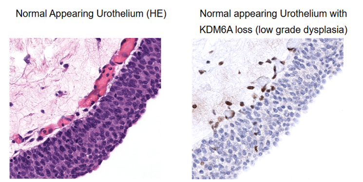
KDM6A (Lysine demethylase 6A, syn. UTX) is an epigenetic regulator which is frequently mutated in urothelial carcinoma. Because KDM6A is located on chromosome X and most of the KDM6A mutations are truncating, most KDM6A mutations result in a complete loss of protein expression and thus become detectable by IHC.
In their study, Viehweger et al. (1) described a KDM6A expression loss in 124 of 345 (35.7%) of pTaG2 low grade, 35 of 152 (23%) of pTaG2 high grade, 17 of 92 (18.5%) of pTaG3, and 225 of 1.128 (19.9%) of pT2-4 urothelial carcinomas of the urinary bladder. As the study was on tissue microarrays and negative samples without positive internal control cells were excluded, the true numbers may even be somewhat higher.
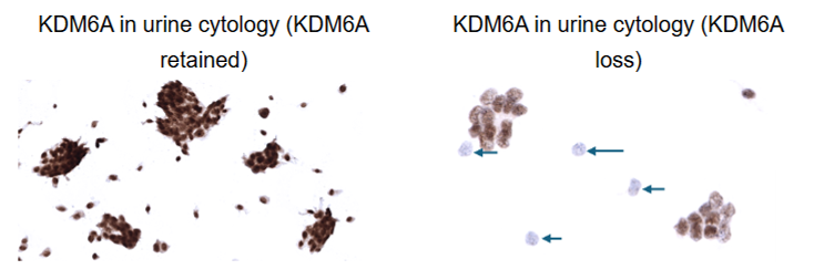
Because of the high-level of KDM6A expression in normal cells and the complete absence of KDM6A expression in affected tumour cells, KDM6A expression losses may also be detectable in urine specimen.
More than 170 further antibodies with a similar level of documentation are provided by MS Validated Antibodies, available to the UK market through 2BScientific.