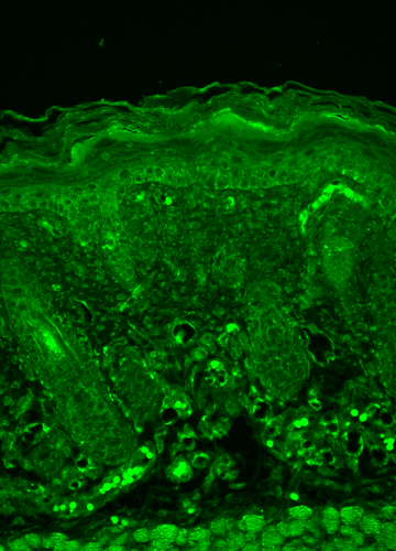
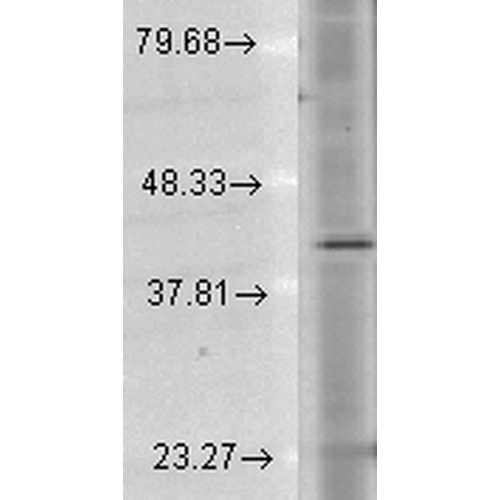
Rhodopsin Antibody, Clone 1D4: Biotin
Product Sizes
100 ug
SMC-177D-BI-100UG
About this Product
- SKU:
- SMC-177D-BI
- Additional Names:
- OPN2, opsd, opsin 2, opsin 2 rod pigment, opsin2, RHO, RP4, MGC138309, Retinitis Pigmentosa 4
- Application:
- ELISA, Immunocytochemistry, Immunofluorescence, Immunohistochemistry, Immunoprecipitation, Western Blot
- Buffer:
- 136.36mM Ethanolamine, 133.23 mM Chlorides, 9.55mM Phosphates, 9.55mM Sodium Bicarbonate
- CE/IVD:
- RUO
- translate.label.attr.clone:
- 1D4
- Clonality:
- Monoclonal
- Conjugate:
- Biotin
- Concentration:
- 1 mg/ml
- Extra Details:
- Rhodopsin is a light-sensitive G protein-coupled receptor (GPCR) found in the rod photoreceptor cells of the retina. It plays a central role in the phototransduction cascade, converting light into electrical signals essential for vision in low-light conditions. Beyond its classical role in visual processing, rhodopsin has emerged as a key player in retinal neurodegeneration. Mutations in the RHO gene encoding rhodopsin are a leading cause of autosomal dominant retinitis pigmentosa (adRP), a progressive neurodegenerative disorder characterized by photoreceptor cell death and vision loss. Misfolded rhodopsin accumulates in the endoplasmic reticulum, triggering cellular stress responses, including the unfolded protein response (UPR) and apoptosis. Rhodopsin dysfunction also affects mitochondrial dynamics, oxidative stress regulation, and autophagy-pathways commonly implicated in broader neurodegenerative diseases such as Alzheimer's and Parkinson's. Its involvement in protein misfolding and trafficking makes rhodopsin a valuable model for studying proteostasis and neurodegeneration. As a well-characterized GPCR with known structural and functional properties, rhodopsin serves as a powerful tool for investigating the molecular mechanisms underlying neuronal survival, degeneration, and therapeutic intervention strategies in neurodegenerative research.
- Host:
- Mouse
- Immunogen:
- Bovine Rhodopsin
- Isotype:
- IgG1
- Purification:
- Protein G Purified
- Reactivities:
- Wide/Non-species Specific
- Shipping Conditions:
- Blue Ice
- Specificity:
- Detects ~40kDa. Recognizes Rhodopsin (native and recombinant forms). No known reactivity to other proteins. Binds specifically to the C-terminal epitope -T-E-T-S-Q-V-A-P-A-(COOH).
- Storage Conditions:
- See Manual
- Supplier:
- StressMarq Biosciences
- Type:
- Antibody: Monoclonal Antibody