Enzo Life Sciences
Enzo Life Sciences has been a leader in innovation and product development to support a diverse range of needs in biomedical research and healthcare for over 45 years.
About Enzo Life Sciences
With a comprehensive portfolio of 20,000 high-quality products including antibodies, genomic probes, assays, biochemicals, and proteins, Enzo supports the work of academic research centers and industry partners who are shaping the future of healthcare around the world.
The company’s proprietary products and technologies play central roles in all areas of translational research and drug development, including genomic analysis, protein analysis, cellular analysis, tissue analysis, and small molecule chemistry.
Enzo has a broad and deep intellectual property portfolio, with patent coverage across a number of key enabling technologies.
AMPIVIEW™
RNA Probes are a cutting-edge tool in the realm of molecular biology and cellular research. At their core, these probes are specialised nucleic acid molecules that are designed to target and bind specifically to particular RNA sequences within cells and tissues. This technique, known as in situ hybridisation (ISH), enables scientists to gain profound insights into gene expression patterns, cellular localisation, and RNA distribution, ultimately enhancing our understanding of various biological processes.
AMPIVIEW® Advantages
- Optimised ready-to-use probe solution for easy usage.
- Considerably more affordable system, saving costs.
- Highly sensitive and specific results you can trust.
- Flexible and faster workflows, suitable for both automated and manual processes
- Compatibility with multiple sample types, offering versatility.
- Preservation of tissue morphology, making it useful for multiplexing and marker detection
- Supported by a range of Enzo's technologies, including enhanced detection reagents like POLYVIEW® PLUS and SAVIEW® PLUS, a variety of chromogens such as HIGHDEF® chromogens, and dedicated wash and hybridisation buffers.
- Custom probes available on request, allowing for specific target detection.

Advanced design & chemistry
What sets AMPIVIEW™ RNA Probes apart from their competitors is their advanced design and chemistry. These probes are engineered to provide exceptional sensitivity and specificity.
Their high degree of specificity ensures that they exclusively bind to the RNA sequences of interest, minimising false-positive signals and background noise.
This specificity is crucial in accurately identifying and studying RNA molecules within complex biological samples.
AMPIVIEW™ RNA Probes excel due to advanced chemistry and design, offering superior specificity, sensitivity, and stronger signal-to-noise ratios compared to older, less specialised FISH technologies.
This makes AMPIVIEW™ a better choice for accurate and reliable RNA detection and localisation in addition to 2BScientific ensuring that you have everything you need for your valued research.
Compatibility with existing ISH setups and protocols
What sets AMPIVIEW™ apart is its compatibility with existing ISH setups and protocols, streamlining the transition for end-users.
These RNA probes find application in a wide spectrum of fields, ranging from basic research and drug discovery/development to clinical research.
Researchers can utilise them for physiological transcription analysis, disease model studies, biomarker research, and target identification and validation.
Moreover, they are invaluable for early preclinical drug discovery analysis, understanding therapeutics' cell response, and advancing companion diagnostic research and development.
The product also caters to the emerging field of Spatial Biology, offering a comprehensive solution for diverse research needs.
Stronger signal-to-noise ratios
AMPIVIEW™ RNA Probes excel due to advanced chemistry and design, offering superior specificity, sensitivity, and stronger signal-to-noise ratios compared to older, less specialised FISH technologies.
This makes AMPIVIEW™ a better choice for accurate and reliable RNA detection and localisation in addition to 2BScientific ensuring that you have everything you need for your valued research.
Enzo p16 and AMPIVIEW™ HPV High-Risk RNA Probes
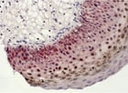
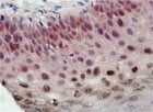
Detection of p16 (red) and HPV High-Risk nucleic acid (brown) detected at the same time in one tissue specimen with Enzo p16 antibody (ENZ-ABS707) and AMPIVIEW™ HPV High-Risk (Dig) RNA Probes detected with POLYVIEW® PLUS AP/HRP and HIGHDEF® Red AP and DAB IHC chromogens.
LoopRNA™ ISH
LoopRNA™ ISH Technology, a proprietary platform developed by Enzo, represents a breakthrough in Molecular Biology and Diagnostics. It plays a pivotal role in elevating the sensitivity of RNA probes, making it a seamless fit within Enzo's AMPIVIEW™ RNA probe portfolio.
These AMPIVIEW™ RNA probes, bolstered by LoopRNA™ ISH technology, offer unparalleled capabilities. They enable the visualisation of nucleic acids at the single-cell level, rivaling the sensitivity of PCR.
These probes provide exceptional specificity while offering design flexibility, catering to various DNA and RNA targets. Detection of AMPIVIEW™ RNA probes is facilitated through Biotin or Digoxigenin loops, easily identifiable using Enzo's dedicated detection reagents like SAVIEW® PLUS and POLYVIEW® PLUS.

MITO-ID® Membrane Potential Kit
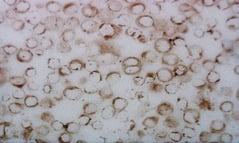
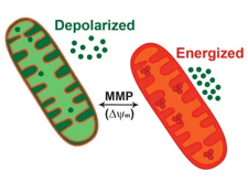
The MITO-ID® Membrane Potential Kit offers a highly sensitive real-time assay for monitoring mitochondrial membrane potential (MMP), crucial in early apoptosis detection.
The kit employs a unique probe that transitions from green fluorescence in the cytosol to orange fluorescence in mitochondria.
The assay is straightforward, requiring no wash steps, and is ten times more sensitive than JC-1, enabling the detection of low drug/dose toxicity without solvent artifacts. With over 50 literature citations, it's a trusted tool for mitochondrial research.
CELLESTIAL® Probes from Enzo Life Sciences
CELLESTIAL® is an advanced live cell dye that revolutionises cell biology research. Its superior photostability guarantees consistent fluorescent signals throughout extended experiments, providing researchers with reliable and reproducible results.
CELLESTIAL® surpasses fix and perm methods with its superior photostability, seamless integration with common dyes, and proven reliability, providing researchers with enhanced precision and consistent results for organelle detection studies in cell biology research. This innovative dye is highly versatile, seamlessly integrating with common dyes and markers like GFP, facilitating multiplex analysis.
CELLESTIAL® is optimised for various platforms, including microplates, flow cytometry, and fluorescent imaging, streamlining workflows for researchers. Its proven track record in peer-reviewed literature makes it a trusted and indispensable tool for scientists striving to advance their understanding of cell biology.
Whether you're studying nuclei, nucleoli, mitochondria, lysosomes, Golgi apparatus, or the endoplasmic reticulum, CELLESTIAL® complements your organelle detection studies, enhancing the precision and reliability of your experiments.
Our complete Enzo range
Please review Enzo's complete range of products designed to meet an incredible breadth of research requirements and be sire to make use of filters identify the most suitable product for our valued customers.

Guidelines
Here you can find all the Enzo Life Science guidelines
-
Enzo Life Science Antibody Guidelines
These guidelines are general tips for use with handling antibody solutions. Antibodies require careful handling procedures to preserve antibody function, stability, and integrity. Please refer to the data sheet(s), product webpage, and Certificate of Analysis for information on individual antibodies.
In the absence of written guidance, contact Technical Support for recommendations on handling of specific proteins.
• Wear appropriate laboratory gear when handling antibodies.
• Prior to opening, centrifuge vials at 10,000 x g for 20 seconds to pull down solution on the sides and top of the vial.
• To avoid contamination and freeze-thaw of your antibody, aliquot the antibody into low-protein-binding-microcentrifuge tubes based on typical experimental usage (no smaller than 10 μL) and freeze at -20 °C or -80 °C based on storage guidelines for your product. Be mindful to place antibodies toward the back of the freezer to avoid temperature fluctuations.
• Conjugated antibodies are particularly vulnerable to freeze thaw cycles. Therefore, many of them are stored at +4 °C while some can be stored at -20 °C. Paying careful attention to their recommended handing is vital to the stability of conjugated antibodies.
• Antibody conjugated to fluorochromes, enzymes or biotin should be stored in dark vials or wrapped in foil as exposure to light will compromise the activity of conjugates.
• Unlabelled, lyophilized antibodies are stable at ambient or 4 °C. Do not freeze unless you are specifically instructed to do so.
• The vial should be tightly capped at all times when not in use.
• Do not use a diluted antibody stored at 4 °C for more than a day. Once diluted, antibodies will become inherently less stable.
• If an antibody must be reconstituted, be sure to use the recommended reconstitution solution and concentration.
• If you are using an antibody for an application for which it has not been recommended, then extensive optimization will be required. Be sure to include appropriate controls, specifically negative controls that do not express your target protein and positive controls which are not part of your experiment sample population (e.g. a recombinant protein or a cell lysate control). -
Enzo Life Science Protein Guidelines
These guidelines are general tips for use with handling proteins. Proteins require careful handling procedures to preserve protein function, stability, integrity, and prevent unwanted protein aggregation. Please refer to the data sheet(s), product web page, and Certificate of Analysis for information on individual proteins.
In the absence of written guidance, contact Technical Support for recommendations on handling of specific proteins.
• Wear appropriate laboratory gear when handling proteins.
• Always centrifuge vial prior to opening to ensure that all protein material has been gathered towards the bottom of the vial.
• Some lyophilized proteins may be difficult to visualize in the tube. Appearance post-lyophilization will vary depending on the protein and the lyophilization buffer/methods used.
• Use the recommended reconstitution solution and concentration.
• The vial should be tightly capped at all times when not in use.
• It is usually best to work with protein solutions on wet ice. Lower temperatures help to retain protein activity levels and can help to prolong protein stability.
• Never vigorously agitate a protein sample. The preferred method of mixing protein solutions is to gently vortex or mix with a micropipette using a polypropylene tip.
• Usually, a quick freeze and a quick thaw are the best methods for retaining protein activities. Avoid repeated freeze-thaw cycles. If a protein solution needs to be frequently taken from the stock material, then it is recommended to aliquot the stock protein material for future use after the initial thaw/reconstitution of the protein.
• For freezing proteins, clearly labelled polypropylene tubes are recommended for snap freezing. Transfer tubes to a pre-chilled box then place tubes in a -80 °C freezer.
• In the absence of a carrier protein, most proteins are unstable at very dilute concentrations. If stability in solution has not been verified, it is best to not dilute carrier-free solutions to protein concentrations below 100 μg/mL unless you intend to use the protein immediately. -
Enzo Life Science - Small Molecules Guidelines
These guidelines are general tips for use with handling small molecules. Small molecules require careful handling procedures to preserve their integrity and stability as well as reduce the chances of contamination and mislabelling. Please refer to the data sheet(s), product webpage, and Certificate of Analysis for information on individual products.
In the absence of written guidance, contact Technical Support for recommendations on the handling of specific small molecules.
• Wear appropriate laboratory gear when handling products.
• Most small molecules are not damaged by multiple freeze-thaw cycles.
• Many of our products are sold in very small quantities, and some are transparent, and/or lyophilized to thin films on the walls of the vials making it very difficult to visually see in the vial.
• We recommend using anhydrous solvents whenever working with compounds that are not water soluble. • Some compounds are difficult to dissolve and will require additional mixing methods such as vortexing, ultrasonication, or heat to get them completely into solution.
• Using the recommended solvents and stock concentration is important. Using our product outside of what is recommended or using concentrations higher than what is suggested could result in solubility issues.
• The vial should be tightly capped at all times when not in use.
• Be advised that small molecules dissolved in DMSO can permeate the skin. Therefore, it is very important to take extra care to avoid skin contact -
Antibodies FAQs
When isolating specific cells by FACS, what concentration of HAT should be added to my culture flasks after fusion?
100uM hypoxanthine, 0.4uM aminopterin, and 16uM thymidine.
Is it possible to perform two-colour detection by staining E6/E7 mRNA and labelling another protein with Alexa Fluor 594 conjugated antibody with ENZ-300?
We have not validated two-colour detection by staining E6/E7 mRNA and labelling another protein with Alexa Fluor 594 conjugated antibody. The E6/E7 RNAs are not labelled, they are hybridized with a labelled probe. The fixation step could cause some problems for the further analysis by PCR, but in general it should be OK.
Liver lysate samples when probed with HO-1 polyclonal antibody, ADI-SPA-896, shows two bands on western blot. What is the possible explanation as two why two bands were detected?
Western blot image displayed on product page with ADI-SPA-896 shows two bands being detected. One of them is full length HO-1 and the lower band is a truncated version. Protein truncations could be the result of protease activity or premature termination due to the presence of a termination codon.
What controls are included in the AMPIPROBE® Bacterial Vaginosis Extraction Controls?
The included controls are as follows:
- Negative Extraction Control
- BV Positive Extraction Control
- No BV Target Control
What is the concentration of α-Gal Epitope (Galα1-3Galβ1-4GlcNAc-R) monoclonal antibody (M86, ALX-801-090-1?
The α-Gal epitope monoclonal antibody is offered as partially purified ammonium sulfate precipitate tissue culture supernatant. The volume provided is 4ml per vial. Since this preparation is not affinity purified mAb, the actual IgM concentration is not determined. To ensure that every lot has a similar amount of mAb, an ELISA is performed to confirm antibody titers.
How is Melanoma marker (human) monoclonal antibody (HMB45), ENZ-C34930 purified from ascites?
Following ammonium sulphate precipitation of ascites, supernatant is further purified on Protein G column.
Please provide the amino acid sequence of the FLAG tag for Irisin (human), (recombinant) (active), ADI-908-307.
FLAG is a short 8 amino acid sequence DYKDDDDK and there is no other linker. Amino acid 32-143 of Irisin comprising the extracellular domain of FNDC5 is fused at the N-terminus to a FLAG®-tag.
What is the lot-to-lot variation for CHO HCP ELISA kit?
In general, the antibodies are stable and we don't change the source of serum/bleeds on a regular basis. Antibodies are purified from the same lot of serum every 3-4 years. Bridging studies have shown they are comparable and there is minimal variation. If there is any slightest detectable lot-to-lot variation in antibody titers, this is self corrected by HCP standards which do not change.
Can a rough estimate antibody concentration of α-Gal Epitope (Galα1-3Galβ1-4GlcNAc-R) monoclonal antibody (M86, ALX-801-090-1 be stated?
The supernatant of ammonium sulfate precipitate hybridoma tissue culture supernatant is concentrated and this can contain up to total protein >10mg/ml. This would amount to approximately less than 5% is the antibody in the preparation by weight.
Is the coated mouse mAb raised against the whole molecule of human FSH ELISA kit?
Coated antibody on FSH Microtiter Plate was raised against complete human FSH.
What is the specific of Detection Ab in FSH ELISA kit?
The detection antibody was raised against the FSHβ subunit. So, beta FSH is what is being detected and measured in this sandwich assay. -
Assays and Kits FAQs
How can I improve the sensitivity of the signal?
Conditions in terms of cell density, culture medium, and incubation have to be respected. It is very important that there is no Cell Growth Medium in the 96 well plate. Presence of Cell Growth Medium can lead to inappropriate conditioning of the cells and could explain lower signals. Cells in Cell Growth Medium have to be washed in PBS and added at the right cell density in Cell Assay Medium to the 96-well plate. The other factor to take into account is the complete reconstitution of Wnt3a in deionized water. Once reconstituted, Wnt3a can be diluted in Assay Diluent. The optimal concentration of Wnt3a has to be determined by the end user with concentrations ranging from 150 to 500ng/mL. Incubation of cells within Wnt3a can be increased from 12 hours to 20 hours if necessary. We also suggest the use of black 96-well plates with clear bottoms to limit well-to-well cross-talk and improve signal-to-noise ratio.
For the Protein A EIA kit, would residual wash buffer affect the assay?
It is best to tap the plate after washing the plate in order to ensure that there is no residual was buffer left in the wells. This can be done gently by tapping the washed plate a couple of times onto a lint free paper towel. It is important to tap for a couple of seconds as to avoid drying the bound material.
Is the LEADING LIGHT Wnt reporter cell line stable?
The LEADING LIGHT Wnt reporter cell line is a stably transfected cell line. Although stable, the cell line will start losing signal between 15 and 20 passages depending on how careful you are with the culture of these cells (maximum of 60% confluence). That is why we generally advise our customers to freeze as many aliquots from early passages as possible. Proceeding this way should allow you to use these cells for a very long period of time and do as many experiments as possible.
For the Protein A EIA kit, would the readings be affected if the wells were to dry out when residual was buffer is blotted out?
Care should be taken to not allow the well to dry out as it could affect the readings.
Can I use the cells straight from the liquid nitrogen?
We would advise against using the cells from the liquid nitrogen. Cells need to be in their exponential growing phase to give relevant results. Furthermore, this cell line is stably transfected and requires a selective environment. Cell Growth Medium contains selective agents. The cells need to go through a phase in Cell Growth Medium prior to conducting the assay. This selection is necessary to guarantee that 100% of the cells in the assay are positive for our luciferase-Wnt responsive element construct.
How long should I incubate my cells after adding the luciferase substrate?
Signals should be read 10 minutes after adding luciferase substrate with a multichannel pippette. The signal is stable enough to add substrate to an entire plate before reading. More than 20-25 minutes and the signal will start to decrease.
Which instrument should I use to measure Wnt activity?
A plate luminometer capable of reading glow chemiluminescence is required. The light emitted by our LEADING LIGHT Wnt reporter cell line has a wavelength of 560nm. Some radiation counters may also be suitable. We typically use a 0.1 second integration time. If it is very weak, you can go up to one second.
Why is 4P curve fit curve recommended for ELISA data analysis and where can I find access to this data analysis software?
The 4P logistic curve fit is a more accurate method for data analysis. Many plate readers do have the ability to perform a 4P curve fit analysis and the software is pre-loaded to the reader/computer. Check with your plate reader manufacturer to confirm if this plate reader can access and use the software.
FAQ's
-
Small molecule FAQs
General handling, storage instructions and stability
Compound libraries should be thawed at room temperature. Once thawed, the plates can be gently centrifuged to bring down any liquid from tops/sides of wells. The cap strips should be carefully removed. You can then aliquot the compounds to additional "daughter" plates if desired. The stability of our libraries very much depends on the number of freeze-thaw cycles. Our compound libraries can be frozen and thawed approximately 6 to 8 times with little or no loss of compound integrity. The master plate and the daughter plates should be stored at -80C. Stock solutions should be stable for at least one year from the date of delivery. Solutions diluted with 100% pure, anhydrous DMSO should also be stored at -80C and may be kept for a least three months. We cannot, however, guarantee the stability of our libraries if you decide on using a different diluent than DMSO to prepare diluted stocks.
Which plates are used to package these libraries?
The libraries are packaged using deep-well 96-well plates from either Corning Costar or Thermo Matrix. The name of the manufacturer is moulded into the plastic at the edge of the plate.
What are compound libraries used for?
In general terms, a laboratory or company interested in high-throughput screening for drug discovery will have already established an assay for the enzyme or system of interest to them. This might be, for example, an enzyme with an activity associated with a particular disease state, or a receptor they believe to be involved in particular signalling cascade or a newly discovered enzyme that they wish to characterise. There is essentially no limit to the number of populations of compounds at the same time. Very often this process is automated robotically to allow for faster processing without the risk of human error. It is completely up to the customer to develop/test/troubleshoot the assay for their system of interest. Once the assay system has been established, the screening process can actually begin. Multiple reaction wells will be set-up (often in a 96-well format, although 384 well-plate can be used). An individual compound from the library is then added to each reaction well. Depending on the assay type, the read out is further assessed by comparing treated with controls. Compounds that either increase or decrease the signal (depending upon the experiment in question) would be considered "hits" or "lead candidates". Following initial screening, lead candidates are further evaluated with additional assays. This could involve assays with purified enzymes to determine inhibitor constants, kinetic studies etc.
Can I customize the library I am interested in?
Libraries can be customized to fit your experimental requirements.
Can I have the IC50 value of the compounds included in your SCREEN-WELL compound libraries?
We do not test for IC50 values. IC50 values are not always available and when they are, they apply to a specific assay under specific conditions.
How are the plate maps and documentation for each compound in libraries provided?
Comprehensive documentation for each compound and the plate map are shipped along with the library on a CD.
Can microcystins be diluted serially into lower concentration working standards with ultrapure water or only with the solvent used to dissolve initially?
Diluting to lower concentrations with solvent should be fine. It is recommended to perform serial dilutions with solvents and not ultra pure water.
Enzo Life Sciences Scientific Posters
Here to help you
At 2BScientific, we follow the motto “No-one does more for the customer” and we look forward to proving that to our valued customers!