Mouse tissue-resident macrophages
Function of tissue-resident macrophages
Tissue-resident macrophages have specialized functions based on their locations and distinct gene expression profiles. They have been implicated in tissue development (e.g. bone formation, brain development, mammary gland development), tissue homeostasis (e.g. maintenance of insulin sensitivity, thermogenic gene induction, hepatic lipid homeostasis) and the resolution of inflammation. In contrast, recruited monocytes - that become macrophages in tissues - are viewed as key players during inflammation and pathogen challenge (Italiani et al., 2014).
Factors shaping the identity of tissue-resident macrophages
Tissue-resident macrophages are a heterogeneous population of immune cells, that fulfill tissue-specific and niche-specific functions (Davies et al., 2013). The phenotype of tissue-resident macrophages is shaped by different factors, that can be subdivided into origin, intrinsic factors and extrinsic factors (figure 1) (Blériot et al., 2020).
Origin. - Tissue-resident macrophages comprise cells arising from three distinct waves of precursors, embryonic yolk sac (YS), fetal liver and adult bone-marrow (BM) derived monocytes with tissue-specific proportions (Blériot et al., 2020). YS-derived macrophages (CD45+ CX3CR1bright F4/80bright) circulate in the blood and colonize the developing mouse embryo between E9.5 and E10.5, starting with the cephalic area. Only from E12.5 onwards when fetal liver hematopoiesis becomes active, a second population of F4/80low CD11bhigh cells appears (Schulz et al., 2012).
These fetal liver derived macrophages largely replace the early YS-derived macrophages, with exception of microglia and a proportion of Langerhans cells, that remain of yolk sac origin (Hoeffel et al., 2015).
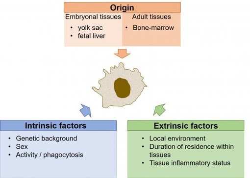
Figure 1: Factors shaping the identity of tissue-resident macrophages (adapted from Blériot et al., 2020).
Murine tissue-resident macrophages derived from embryonic precursors, maintain their numbers in adults by self-renewal (Ginhoux et al. 2016). A third tissue-resident macrophage population arises from adult BM-derived precursors. As a consequence, each adult tissue contains a mosaic of three ontogenically distinct macrophage populations.
Intrinsic factors. - Intrinsic factors affecting tissue-resident macrophage identity include the genetic background and the sex. Mutations in myeloid genes are associated with diseases such as myeloid leukemia (Mueller et al., 2002). Sexual identity influences for example microglial properties (Thion et al., 2018).
Extrinsic factors. - How extrinsic factors are shaping the identity of tissue-resident macrophages is only just started to be identified. The local environment or "niche of residence" has a major impact on the macrophage phenotype. As tissues are not uniform - they consist of different cell types - organ-specific macrophages are also not uniform. Instead, within the same tissue different programs may be present in different subpopulations of macrophages located in unique sub-tissular niches (Blériot et al., 2020). The time that macrophages spent in a specific local environment furthermore impacts the macrophage identity. Newly arrived monocyte derived macrophages can co-exist with embryonic derived macrophages within the same organ.
Markers for murine tissue-resident macrophages
Murine tissue-resident macrophages derived from yolk sac cells are characteristically F4/80high macrophages, e.g. Langerhans cells in the skin, Kupffer cells in the liver and microglia in the brain (Schulz et al., 2012). F4/80low CD11bhigh macrophages are of hematopoietic origin and are continuously replaced by BM derived progenitors. BM progenitors replace also classical dendritic cells and some F4/80high macrophages found particularly in the kidney and the lung (Schulz et al. 2012). Regardless of their origin, genetic and cell culture studies indicate the major lineage regulator of almost all macrophages is the colony stimulating factor 1 receptor, CSF1R (Wynn et al., 2013).
Table 1: Markers of murine tissue-resident macrophages available from SYSY / HistoSure.
| Marker | Antibody | Application |
| CD 11b | HS-384 103, rabbit, pAb HS-384 117, rat, mAb | Frequently used to identify macrophages and microglia, however not expressed by all tissue-resident macrophages. |
| CD 11c | HS-375 003, rabbit, pAb HS-375 004, Guinea pig, serum | Expressed on dendritic cells, monocytes, macrophages, neutrophils and a small subset of B-cells. |
| CD45 | HS-427 017, rat, mAb | A marker protein for murine leukocytes. Expression level of CD45 is used to identify macrophage subpopulations. |
| CD163 | HS-455 003, rabbit, pAb | Mainly expressed by tissue-resident macrophages. Marker of perivascular macrophages in the CNS. |
| Chil3 | 442 004, Guinea pig, serum | Secreted protein primarily produced by macrophages during inflammation. |
| F4/80 | HS-397 004, Guinea pig, serum HS-397 008, rabbit, mAb HS-397 017, rat, mAb | Marker of murine macrophages. Expression varies between macrophage sub-populations. |
| IBA1 | HS-234 004, Guinea pig, serum HS-234 013, rabbit, pAb | Marker specific for microglia and macrophages. |
| Tmem 119 | 400 008, rabbit, mAb 400 004, Guinea pig, serum | Highly selective microglia marker protein. |