FluoTags
FluoTags are camelid single domain antibodies consisting only of one antigen binding site of a Alpaka heavy chain antibody. With only ~15 kDa, these Tags are about 10-times smaller than conventional IgG antibody molecules.
In FluoTag-Q each fluorophore is coupled to exactly one FluoTags, which in turn binds to its target molecule in a monovalent fashion. The high binding affinity and a coupling efficiency of > 95% guarantees a highly linear relation between the number of target molecules and the intensity of fluorescence. This enables a direct count of the target molecule of interest. The fluorophore is located exceptionally close to the recognized epitope (less than 1.5 nm), which is ideal for all microscopy techniques.
In FluoTag-X two fluorophore molecules are site-specifically coupled to each FluoTags molecule. Therefore, the reagent simultaneously targets up to four fluorophores to the protein of interest, which ensures extra-bright signals. Owing to the small size of the FluoTags, the distance between the target epitope and each fluorophore is below 4 nm.
In comparison to detection systems using conventional antibodies, FluoTag-X can thus improve the localization accuracy by 10-15 nm. Both features - superior brightness and precise fluorophore placement - render the FluoTag-X products excellent tools for all microscopy techniques.
Advantages:
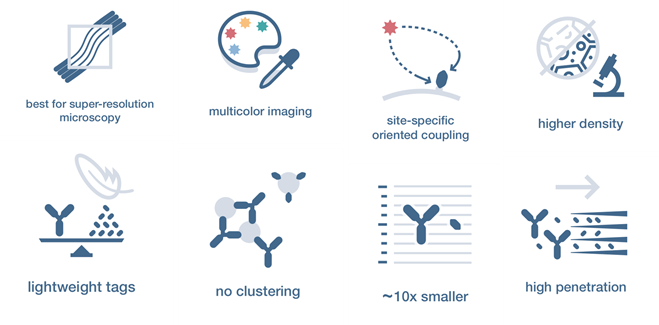
10x Smaller
With only ~15 kDa, our Tags are about 10-times smaller than conventional IgG antibody molecules. This simple but significant difference is the core feature that adds extra benefits to our tools.
High Penetration
Our FluoTags can freely move in crowded environments like fixed tissue or cells. Their fast diffusion allows them to find epitopes faster. They also can reach epitopes that conventional IgGs have no access to.
High Labeling Density
Steric hindrance is minimal with our FluoTags. This means that FluoTags will find more epitopes than conventional IgGs. This results in a more uniform decoration of the structures under investigation.
No Clustering
Staining of live or poorly fixed samples is often prone to clustering artifacts. FluoTags are monovalent and recognize only one epitope. They therefore efficiently prevent clustering of target molecules or organelles.
Multicolor Imaging
We can couple FluoTags to your favorite fluorophore. No need to match the secondary antibody to the species of your primary. In addition, direct coupling can significantly shorten your experimental time.
Site-Specific Coupling
FluoTags contain a fixed number of fluorophores at defined positions. In our FluoTag-Q series, for example, each fluorophor is linked to exactly one FluoTags molecule at a position less than 1.5 nm apart from the epitope
Super-Resolution
FluoTags locate the fluorophores 10-15 nm closer to their targets than classical detection systems. This improves the localization accuracy required for all modern super-resolution microscopy techniques.
Multiplexing
Multiplexing immunofluorescence allows researchers to stain a single sample with multiple primary antibodies originating from the same species, even if they have identical isotype. Staining can, e.g., be performed with multiple monoclonal mouse IgG1 kappa primary antibodies or with multiple polyclonal antibodies raised in rabbit at the same time.
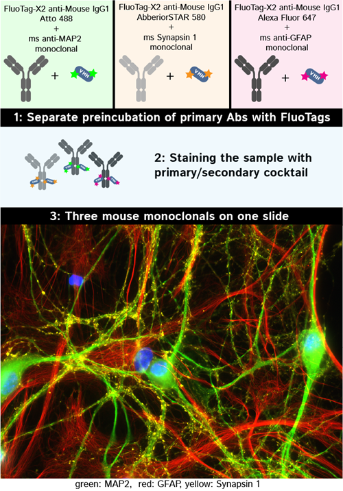
Indirect immunofluorescence labeling of PFA fixed hippocampus neuron with three mouse monoclonals premixed and stained simultaniously. FluoTag-X2 anti-Mouse IgG kLC conjugated to three different fluorophores Atto 488, AbberiorSTAR 580, Alexa Fluor 647 (cat. no. N1202-At488-L, N1202-Ab580-L, N1202-AF647-L) were used to stain Mouse anti-MAP 2 (cat. no. 188 011, green), Mouse anti-GFAP (cat. no. 173 011, red) and Mouse anti-Synapsin 1(cat. no. 106 011, yellow). Nuclei have been visualized by DAPI staining (blue).
Primary antibody requirements
For multiplexing applications, all primary antibodies need to be well characterized and of high quality. As the stoichiometry between primary antibody and the secondary FluoTags reagent is critical, the concentration of the primary antibody needs to be known precisely. Monoclonal antibodies therefore need to be purified e.g. via Protein A to remove serum proteins. Affinity purification is mandatory for polyclonal tools in order to remove serum proteins and antibodies not recognizing the target protein.
Recommended trial experiments
Multiplexing in general is a delicate application, which may require optimization. We strongly recommend to first establish conventional single-color staining using secondary FluoTags reagents in order to get a feeling for the specific properties, blocking requirements and optimal concentration range of the individual primary antibodies used.