Antibodies Against All Desmosome Protein
Desmosomes – mechanical stabilizers in cellular junctions
Desmosome structure with arrangement of major desmosomal components according to Holthöfer et al., 2007[1] (IDP: inner desmosomal plaque, ODP: outer desmosomal plaque).
Desmosomes are important structures in the assembly of cell-cell adhesion, where they establish the mechanical coupling of neighboring cells.1,2,3 A protein complex that is anchored in the intracellular intermediate filament system mediates strong noncovalent interactions between its components and is responsible for the extreme stability of the desmosome unit (see adjacent schematic drawing).1
Not surprisingly, desmosomal proteins are highly expressed in tissues with intense mechanical stress, such as skin or cardiac muscle. Besides their structural function, proteins of the desmosomal plaque play a role in cell communication and signaling, in particular during cell proliferation, cell migration, and cell assembly in tissue (re-)generation.2,3
In cell biology research, desmosome proteins are useful markers for junction formation, disassembly, and differentiation, e.g. in developmental studies. Regenerative biology investigates desmosome function in wound healing or tissue repair.

Role of desmosomes in health and disease
Desmosome malfunction, caused by genetic defects, autoantibodies, or malignancies is involved in
the pathogenesis of diseases that especially affect tissues and organs under intense mechanical stress
(see table below).1,2,3
Mutations of desmosomal proteins have been linked to multiple disorders, such
as cardiomyopathies, autoimmune, or skin diseases. In addition, the role of misregulated proteins in
genesis and progression of many tumors is under intense research.2,3
| Desmosomal component | Genetic / protein alteration | Disease / phenotype |
| desmoplakin | nonsense or missense mutations | ARVC (arrhythmogenic right ventricular cardiomyopathy), epidermolysis bullosa, Carvajal/Naxos syndrome with/without dental phenotype |
| deletion in mouse | tumor invasion in model of pancreatic carcinogenesis and non small cell lung cancer | |
| plakophilin 2 | nonsense or missense mutations | ARVC, Brugada syndrome |
| plakophilin 3 | elevated mRNA | gastrointestinal cancer |
| increased expression | breast cancer, pancreatic cancer | |
| plakoglobin | nonsense and splice site | skin diseases, e.g. skin fragility, palmoplantar keratoderma or |
| conditional mouse knock-out | ARVC | |
| desmoglein 1 | missense mutations and nucleotide deletion | severe skin dermatitis, multiple allergies, metabolic wasting |
| desmoglein 2 | increased expression | malignant skin carcinomas |
Versatile antibody portfolio for all relevant antigens
PROGEN features a comprehensive portfolio of over 20 antibodies against all relevant desmosome constituents, such as desmoglein, desmoplakin, plakoglobin, or plakophilin. The different formats are suitable for a wide range of applications in basic research or pathology, such as immunohistochemistry (IHC), western blot (WB), immunocytochemistry (ICC), or immunofluorescence (IF).
Many IgGs are available from different hosts to enable co-stainings. In addition, PROGEN offers matching secondary antibodies as well as IHC staining kits to facilitate workflows in research and pathology routine. Most desmosome antibodies are available in ready-to-use sample sizes (600 μL serum) and common markers have been combined to convenient, cost-effective sets (see table below).
The desmosome product line is part of PROGEN's cell adhesion research antibody portfolio that includes other central markers of cellular junctions (e.g. cadherin or catenin). In addition, a wide range of cytoskeletal and keratin antibodies are available for comprehensive studies.
Enquire at Info@2BScientific.com
| Test Our Sets! | Antibodies Against | Cat No. |
| anti-desmosome sample set 1 | desmoglein 1, desmocollin 1, plakophilin 1, plakoglobin, and desmoplakin 1/2 | 70030 |
| anti-desmosome sample set 2 | desmoglein 1/2, plakophilin 2, plakoglobin, and desmoplakin 1/2 | 70031 |
| anti-desmosome sample set 3 | desmoglein 3, desmocollin 3, plakophilin 3, plakoglobin, and desmoplakin 1/2 | 70032 |
| anti-desmoglein 1-4 sample set | desmoglein 1, 2, 3, and 4 | 70033 |
| anti-desmocollin 1 & 3 sample set | desmocollin 1 and 3 | 70034 |
| anti-plakophilin 1-3 sample set | plakophilin 1, 2, and 3 | 70035 |
Established premium quality
PROGEN's desmosome antibodies are highly published and have been independently validated for relevant applications. Protocols and data provide useful information for experimental design.
 Desmocollin-1 immunohistochemistry of rat dorsal skin(courtesy of J. Heß, University Hospital Heidelberg)
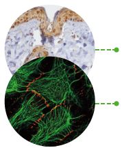
Immunofluorescent double staining of HaCaT cells (red:
plakophilin-1, green: cytokeratin; courtesy of L. Langbein,
University Hospital Heidelberg)
Referenced Reviews
- Holthoefer B et al., 2007, Structure and function of desmosomes, Int. Rev. Cytol., Vol.
264; ISSN 0074-7696, Elsevier Inc. - Broussard JA et al., 2015, Desmosome regulation and signaling in disease, Cell Tissue Res., 360(3): 501–512
- Johnson JL et al., 2014, Desmosomes: regulators of cellular signaling and adhesion in epidermal health and disease, Cold Spring Harb Perspect Med 2014;4:a015297