Mabtech
Mabtech is a Swedish biotech company that was founded in 1986 by scientists at the Department of Immunology, Stockholm University. One of only very few companies worldwide focused entirely on developing monoclonal antibodies for ELISpot, FluoroSpot and ELISA applications, Mabtech supports the study of key immunological targets that include cytokines, immunoglobulins and apolipoproteins.
We rely heavily on Mabtech to provide our customers with highly characterised monoclonal antibodies. These are supplied as individual products, configured as optimal antibody pairs, or included as essential components of a wide variety of user-friendly kits. Product quality is assured through ISO accreditation (ISO 9001 and ISO 13485), as well as through citation in many high impact publications.
Research areas for which we frequently recommend Mabtech’s kits include infectious disease, cancer, allergy, transplantation and vaccine monitoring, and we also endorse Mabtech antibodies for cardiovascular and veterinary research.
Researchers with an interest in immune monitoring regularly turn to us as a source of Mabtech ELISpot kits, especially those who have invested in Mabtech’s IRIS™ ELISpot reader. One question we’re often asked is ‘what are the main differences between Mabtech’s ELISpot, FluoroSpot and ELISA kits?’. While all these products are based on the same high-quality Mabtech antibodies, they differ in terms of configuration.
Mabtech ELISAs are performed within a standard microplate, typically resulting in a colorimetric readout, whereas during a Mabtech ELISpot assay the microplate wells contain a PVDF membrane coated with a capture antibody to quantify cytokine-releasing cells. The FluoroSpot assay builds on the ELISpot principle by providing the capacity to analyze the secretion of multiple analytes from the same cell.
The ELISA assay relies on the antibodies that capture and detect the analyte of target. We at Mabtech put a lot of effort into developing and characterizing the antibodies. The antibody clones are often the same as in ELISpot, a method that requires optimal sensitivity and specificity of the antibodies involved. We always validate the ELISAs for recognition of native proteins as it often differs from how the antibodies bind to the recombinant protein. The use of quality antibodies makes our ELISAs both very specific and sensitive.
Mabtech ELISpot
Cell-based immunoassay with detection levels as low as one cell in a million
ELISpot can be used in almost any setting to investigate specific immune responses including vaccines cancer and diagnostics.
Study analyte secretion by capturing analytes immediately and throughout the process.
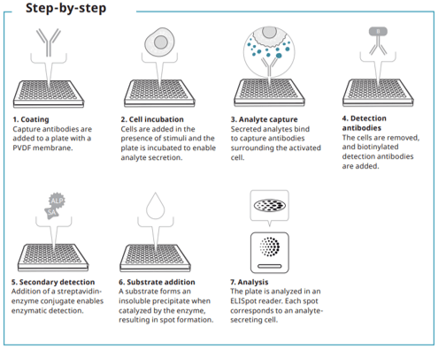
ELISpot is used to quantify protein-secreting cells. In this assay, cells are cultured in a plate coated with capture antibody. Cytokines, immunoglobulins, or other target proteins secreted by the cells are captured immediately after secretion and throughout the stimulation process. After cell removal, secreted proteins are identified using a detection antibody. Visible spots form after adding a precipitating substrate. Each spot corresponds to an individual analyte-secreting cells. ELISpot can quantify analyte secreting cells at the single cell level and is considered one of the most sensitive cellular assays available. Additionally, ELISpot is very easy to perform and is suitable for the analysis of many samples at different timepoints. The assay can be automated using a 96-well plate and analysed rapidly with an automated ELISpot reader.
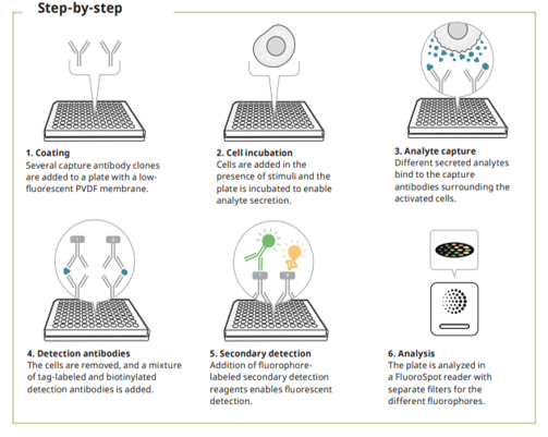
Mabtech Fluorospot
- Multiplex version of ELISpot
- Combine analytes of different kinetics without manipulating intracellular processes
- Robust and easy to scale up from discovery phase to larger studies.
FluoroSpot is used to quantify protein-secreting cells. This immunoassay combines the sensitivity of ELISpot with the capacity to study the secretion of several analytes simultaneously. In FluoroSpot, a cell suspension is added to the wells of a plate. Proteins, secreted by cells are captured immediately after secretion and throughout the stimulation process by specific antibodies. After cell removal, tagged or biotinylated detection antibodies are added, followed by fluorophore-labeled secondary reagents. The captured proteins turn into fluorescent spots, where each spot corresponds to a single cell.