600-401-EA2
RIP1 Antibody

Cannot supply to this region.
- SKU:
- 600-401-EA2
- Additional Names:
- RIP1 Antibody, RIP, RIP1, RIP, Cell death protein RIP, RIP-1|RIPK1
- Application:
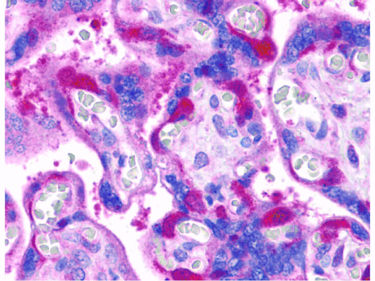
- ELISA, IHC
- Concentration:
- 1 mg/ml
- Physical State:
- Liquid
- Species Reactivity:
- Human
- Storage Conditions:
- -20[o]C aliquoted. Aliquot. Avoid freeze/thaw cycles., 2-8[o]C diluted. Aliquot. Avoid freeze/thaw cycles.
- Supplier:
- Rockland Inc
- Host:
- Rabbit
- Reactivities:
- Human
- Buffer:
- 0.01 M Sodium Phosphate, 0.25 M Sodium Chloride
- Immunogen:
- Anti-RIP1 antibody was prepared from whole rabbit serum produced by repeated immunizations with a 14 amino acid peptide near the C-terminus of human RIP1.
- Formulation:
- 0.01 M Sodium Phosphate, 0.25 M Sodium Chloride, pH 7.2
- Uniprot:
- Q13546
- Synonyms:
- AIEFL;cell death protein RIP;IMD57;receptor (TNFRSF)-interacting serine-threonine kinase 1;receptor-interacting protein 1;receptor-interacting protein kinase 1;receptor-interacting serine/threonine-protein kinase 1;RIP;RIP-1;RIP1;serine/threonine-protein kinase RIP
- Extra Details:
- RIP1 (Receptor Interacting Protein), also known as RIPK1, is a crucial 74 kD adaptor kinase in several of stress-induced signaling pathways and on the crossroad of a cell's decision to live or die. RIP1 contains an N-terminal region with homology to protein kinases, an intermediate domain capable of association with MAPKKK and a C-terminal region containing a death domain motif present in the Fas and TNFR1 intracellular domains. Full length RIP1 is important for signaling to NF-kappa-B, MAPKs and necrosis, whereas caspase-8 generates a C-terminal RIP1 cleavage fragment, promoting TNF-induced apoptosis. It is required for TNFRSF1A-mediated and TLR3-induced NF-kappa-B activation. RIP1-deficient mice fail to thrive, displaying extensive apoptosis in both lymphoid and adipose tissues and dying at 1-3 days of age.
- Shipping Conditions:
- Dry Ice