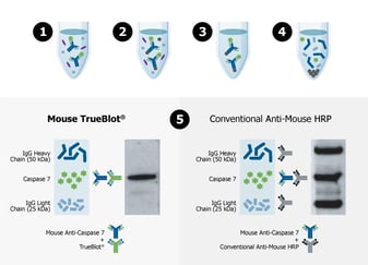
Immunoprecipitation (IP) and Western blot (WB) protocols provide highly specific results but often suffer from poor or non-specific binding, high background, and heavy/light chain interference.
Rockland’s TrueBlot® line of products consists of both TrueBlot® IP beads and TrueBlot® monoclonal secondary antibodies, which combat these common experimental issues by providing increased specificity, low background, and enhanced accuracy to provide publication-quality IP Western blots without interference from contaminating IgG.
The TrueBlot® product line is ideal for studying protein-protein interactions, post-translational modifications (phosphorylation and acetylation), poorly expressed proteins, and immunoprecipitated proteins with molecular weights similar to the heavy and light chain.
TrueBlot® secondary antibodies only recognize primary antibodies in their native (non-reduced) state, eliminating interference from heavy and light chain bands to provide accurate, publication-quality Western blots of immunoprecipitated samples. Pair TrueBlot® antibodies with the exceptional purification properties provided by TrueBlot® IP beads to eliminate the common issues of IP-Western Blot.
Fluorescent TrueBlot® Anti-Rabbit Ig Fluorescein (Cat. No. 18-0216-32)
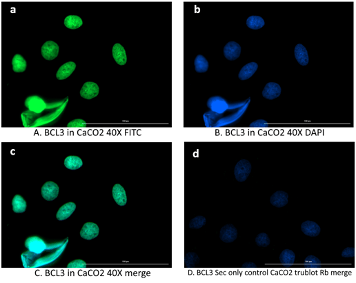
Immunofluorescence microscopy of BCL3 in Caco-2 cells using FITC-conjugated Fluorescent TrueBlot® anti-rabbit Ig for detection.
Caco-2 cells were fixed with 4% PFA, blocked (5% mouse serum/0.3% Triton X-100 in 1X PBS ) for 1 hr, then incubated with 15 µg/mL of anti-BCL3 primary antibody (Cat. No. 600-401-GU4) at 4 °C overnight. Following 3 washes in 1X PBS for 5 min each, 5 µg/mL of FITC-conjugated Fluorescent TrueBlot® anti-rabbit IgG was added and allowed to incubate for 1 hr at room temperature. Nuclei were counterstained with DAPI present in mounting medium. The predicted main localization is nucleoplasm. Additional localization in some cell types includes vesicles and midbody. (a) BCL3 (b) DAPI (c) merged DAPI/BCL3 (d) secondary antibody only. Image taken at 40X magnification.