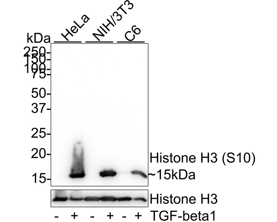
Western blot analysis of Phospho-Histone H3 (S10) on different lysates with Rabbit anti-Phospho-Histone H3 (S10) antibody (ET1601-30) at 1/5;000 dilution. Lane 1: HeLa whole cell lysate (20 µg/Lane) Lane 2: HeLa treated with 50nM Calyculin A for 30 minutes whole cell lysate (20 µg/Lane) Lane 3: NIH/3T3 whole cell lysate (20 µg/Lane) Lane 4: NIH/3T3 treated with 100nM Calyculin A for 30 minutes whole cell lysate (20 µg/Lane) Lane 5: C6 whole cell lysate (20 µg/Lane) Lane 6: C6 treated with 100nM Calyculin A for 30 minutes whole cell lysate (20 µg/Lane) Predicted band size: 15 kDa Observed band size: 15 kDa Exposure time: 10 seconds; 4-20% SDS-PAGE gel. Proteins were transferred to a PVDF membrane and blocked with 5% NFDM/TBST for 1 hour at room temperature. The primary antibody (ET1601-30) at 1/5;000 dilution was used in 5% NFDM/TBST at 4℃ overnight. Goat Anti-Rabbit IgG - HRP Secondary Antibody (HA1001) at 1:50;000 dilution was used for 1 hour at room temperature.
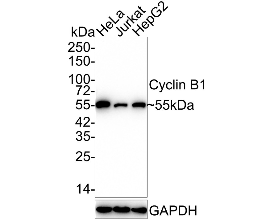
Western blot analysis of Cyclin B1 on different lysates with Rabbit anti-Cyclin B1 antibody (ET1608-27) at 1/5;000 dilution. Lane 1: HeLa cell lysate Lane 2: Jurkat cell lysate Lane 3: HepG2 cell lysate Lysates/proteins at 15 µg/Lane. Predicted band size: 48 kDa Observed band size: 55 kDa Exposure time: 3 minutes 10 seconds; 4-20% SDS-PAGE gel. Proteins were transferred to a PVDF membrane and blocked with 5% NFDM/TBST for 1 hour at room temperature. The primary antibody (ET1608-27) at 1/5;000 dilution was used in 5% NFDM/TBST at 4℃ overnight. Goat Anti-Rabbit IgG - HRP Secondary Antibody (HA1001) at 1:50;000 dilution was used for 1 hour at room temperature.