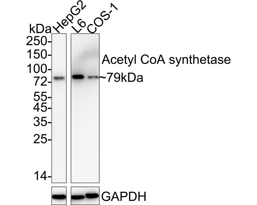
Western blot analysis of Acetyl CoA synthetase on different lysates with Rabbit anti-Acetyl CoA synthetase antibody (ET1702-21) at 1/1;000 dilution. Lane 1: HepG2 cell lysate (20 µg/Lane) Lane 2: L6 cell lysate (20 µg/Lane) Lane 3: COS-1 cell lysate (20 µg/Lane) Predicted band size: 79 kDa Observed band size: 79 kDa Exposure time: 3 minutes; ECL: K1801; 4-20% SDS-PAGE gel. Proteins were transferred to a PVDF membrane and blocked with 5% NFDM/TBST for 1 hour at room temperature. The primary antibody (ET1702-21) at 1/1;000 dilution was used in 5% NFDM/TBST at 4℃ overnight. Goat Anti-Rabbit IgG - HRP Secondary Antibody (HA1001) at 1/50;000 dilution was used for 1 hour at room temperature.
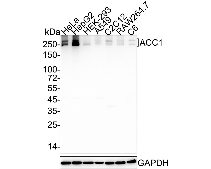
Western blot analysis of Acetyl CoA Carboxylase 1 (ACC1) on different lysates with Rabbit anti-Acetyl CoA Carboxylase 1 (ACC1) antibody (ET1609-77) at 1/2;000 dilution. Lane 1: HeLa cell lysate (20 µg/Lane) Lane 2: HepG2 cell lysate (20 µg/Lane) Lane 3: HEK-293 cell lysate (20 µg/Lane) Lane 4: A549 cell lysate (20 µg/Lane) Lane 5: C2C12 cell lysate (20 µg/Lane) Lane 6: RAW264.7 cell lysate (20 µg/Lane) Lane 7: C6 cell lysate (20 µg/Lane) Predicted band size: 266 kDa Observed band size: 250/266 kDa Exposure time: 43 seconds; 4-20% SDS-PAGE gel. Proteins were transferred to a PVDF membrane and blocked with 5% NFDM/TBST for 1 hour at room temperature. The primary antibody (ET1609-77) at 1/2;000 dilution was used in 5% NFDM/TBST at 4℃ overnight. Goat Anti-Rabbit IgG - HRP Secondary Antibody (HA1001) at 1/50;000 dilution was used for 1 hour at room temperature.
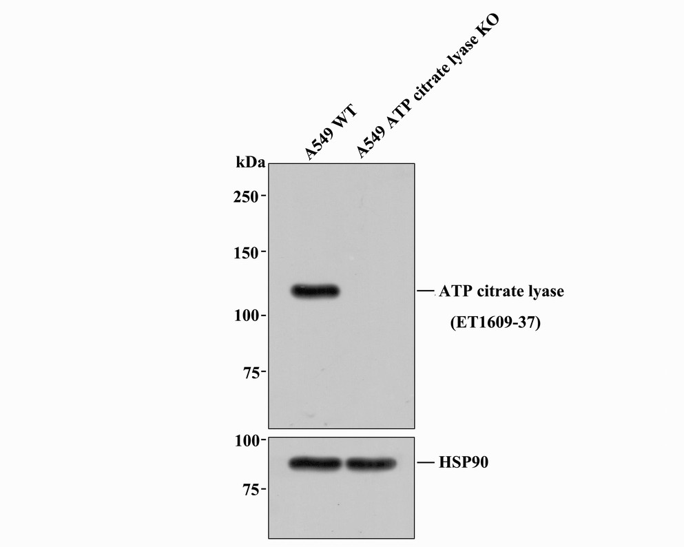
Western blot analysis of ATP citrate lyase with anti-ATP citrate lyase antibody [ST51-07] (ET1609-37) at 1/1;000 dilution. Lane 1: Wild-type A549 whole cell lysate (20 µg). Lane 2: ATP citrate lyase knockout A549 whole cell lysate (20 µg). ET1609-37 was shown to specifically react with ATP citrate lyase in wild-type A549 cells. No band was observed when ATP citrate lyase knockout sample was tested. Wild-type and ATP citrate lyase knockout samples were subjected to SDS-PAGE. Proteins were transferred to a PVDF membrane and blocked with 5% NFDM in TBST for 1 hour at room temperature. The primary antibody (ET1609-37; 1/1;000) and Loading control antibody (Rabbit anti-HSP90; ET1605-56; 1/10;000)was used in 5% BSA at room temperature for 2 hours. Goat Anti-Rabbit IgG-HRP Secondary Antibody (HA1001) at 1:200;000 dilution was used for 1 hour at room temperature.
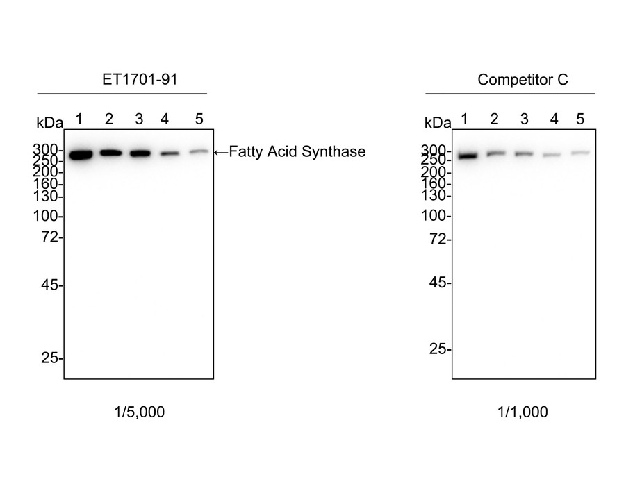
Western blot analysis of Fatty Acid Synthase on different lysates with Rabbit anti-Fatty Acid Synthase antibody (ET1701-91) at 1/5;000 dilution and competitor's antibody at 1/1;000 dilution. Lane 1: HeLa cell lysate Lane 2: HEK-293 cell lysate Lane 3: A549 cell lysate Lane 4: C2C12 cell lysate Lane 5: L-929 cell lysate Lysates/proteins at 20 µg/Lane. Predicted band size: 273 kDa Observed band size: 273 kDa Exposure time: 1 minute 2 seconds; 4-20% SDS-PAGE gel. Proteins were transferred to a PVDF membrane and blocked with 5% NFDM/TBST for 1 hour at room temperature. The primary antibody (ET1701-91) at 1/5;000 dilution and competitor's antibody at 1/1;000 dilution were used in 5% NFDM/TBST at 4℃ overnight. Goat Anti-Rabbit IgG - HRP Secondary Antibody (HA1001) at 1/50;000 dilution was used for 1 hour at room temperature.