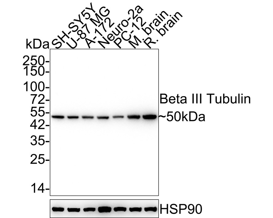
Western blot analysis of Beta III Tubulin on different lysates with Mouse anti-Beta III Tubulin antibody (M0805-8) at 1/2;000 dilution. Lane 1: SH-SY5Y cell lysate Lane 2: U-87 MG cell lysate Lane 3: A-172 cell lysate Lane 4: Neuro-2a cell lysate Lane 5: PC-12 cell lysate Lane 6: Mouse brain tissue lysate Lane 7: Rat brain tissue lysate Lysates/proteins at 10 µg/Lane. Predicted band size: 50 kDa Observed band size: 50 kDa Exposure time: 11 seconds; 4-20% SDS-PAGE gel. Proteins were transferred to a PVDF membrane and blocked with 5% NFDM/TBST for 1 hour at room temperature. The primary antibody (M0805-8) at 1/2;000 dilution was used in 5% NFDM/TBST at 4℃ overnight. Goat Anti-Mouse IgG - HRP Secondary Antibody (HA1006) at 1/50;000 dilution was used for 1 hour at room temperature.
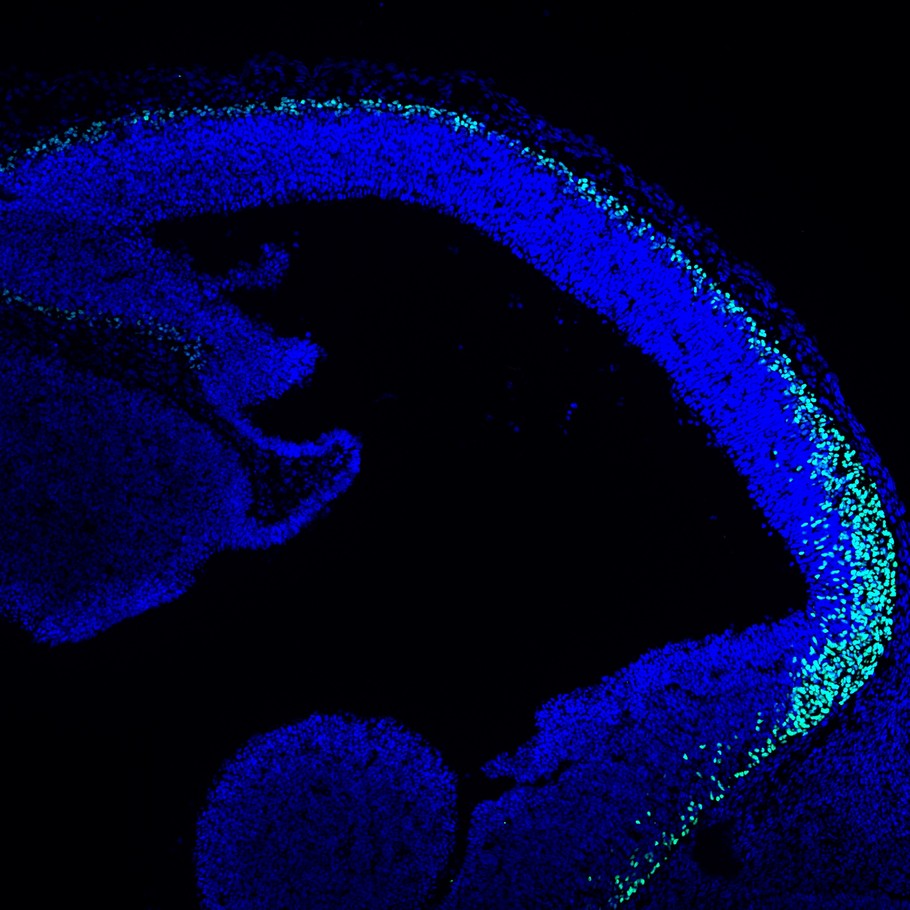
Immunofluorescence analysis of frozen E14.5 mouse embryonic brain tissue with Rabbit anti-TBR1 antibody (ET1702-97) at 1/500 dilution. The section was pre-treated using heat mediated antigen retrieval with sodium citrate buffer (pH 6.0) for about 2 minutes in microwave oven. The tissues were blocked in 10% negative goat serum for 1 hour at room temperature; washed with PBS; and then probed with the primary antibody (ET1702-97; green) at 1/500 dilution overnight at 4 ℃; washed with PBS. Goat Anti-Rabbit IgG H&L (iFluor™ 488; HA1121) was used as the secondary antibody at 1/1;000 dilution. Nuclei were counterstained with DAPI (blue).
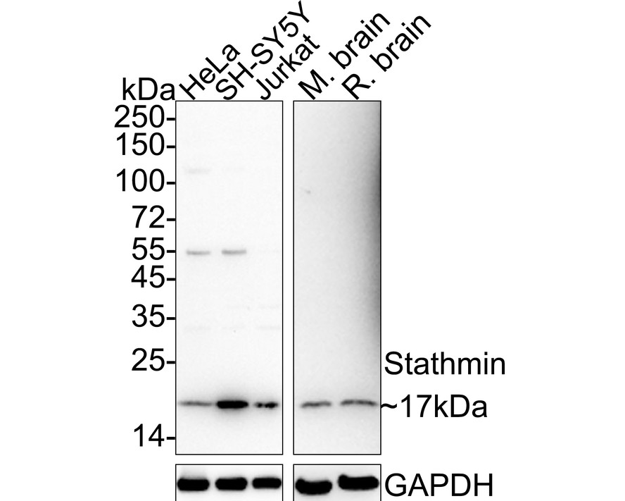
Western blot analysis of Stathmin on different lysates with Rabbit anti-Stathmin antibody (HA721179) at 1/1;000 dilution. Lane 1: HeLa cell tissue lysate (20 µg/Lane) Lane 2: SH-SY5Y cell tissue lysate (20 µg/Lane) Lane 3: Jurkat cell tissue lysate (20 µg/Lane) Lane 4: Mouse brain tissue lysate (40 µg/Lane) Lane 5: Rat brain tissue lysate (40 µg/Lane) Predicted band size: 17 kDa Observed band size: 17 kDa Exposure time: 3 minutes; ECL: K1801; 4-20% SDS-PAGE gel. Proteins were transferred to a PVDF membrane and blocked with 5% NFDM/TBST for 1 hour at room temperature. The primary antibody (HA721179) at 1/1;000 dilution was used in 5% NFDM/TBST at 4℃ overnight. Goat Anti-Rabbit IgG - HRP Secondary Antibody (HA1001) at 1/50;000 dilution was used for 1 hour at room temperature.
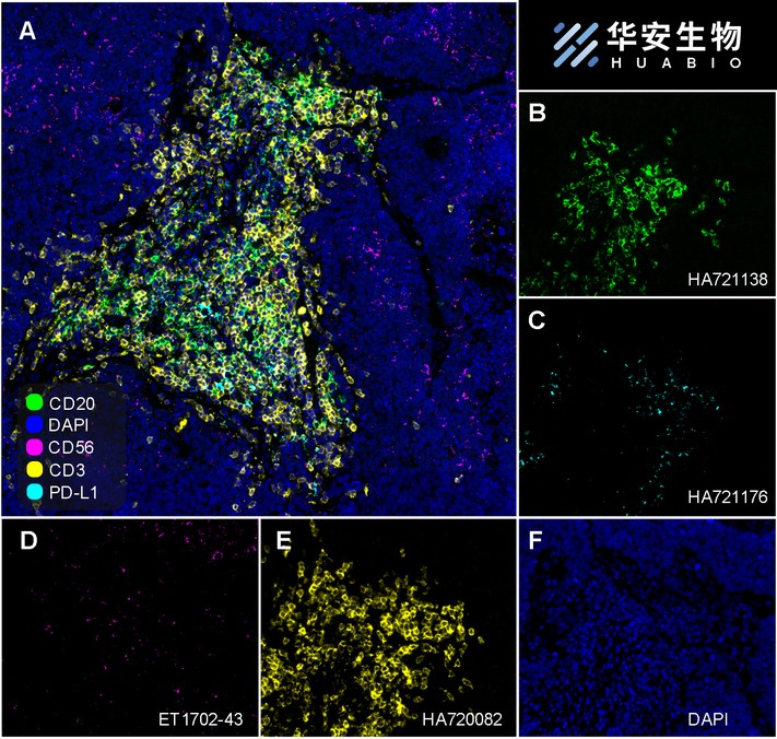
Fluorescence multiplex immunohistochemical analysis of Tertiary Lymphoid Structures in Human Small Cell Lung Cancer (Formalin/PFA-fixed paraffin-embedded sections). Panel A: the merged image of anti-CD20 (HA721138; green); anti-PD-L1 (HA721176; cyan); anti-CD56 (ET1702-43; magenta) and anti-CD3 (HA720082; yellow) on tertiary lymphoid structures. Panel B: anti- CD20 stained on B cells. Panel C: anti-PD-L1 stained on dendritic cells and macrophages cells. Panel D: anti-CD56 stained on NKT cells. Panel E: anti-CD3 stained on T cells. HRP Conjugated UltraPolymer Goat Polyclonal Antibody HA1119/HA1120 was used as a secondary antibody. The immunostaining was performed with the Sequential Immuno-staining Kit (IRISKit™MH010101; www.luminiris.cn). The section was incubated in four rounds of staining: in the order of HA721138 (1/1;500 dilution); HA721176 (1/1;000 dilution); ET1702-43 (1/1;000 dilution); and HA720082 (1/500 dilution) for 20 mins at room temperature. Each round was followed by a separate fluorescent tyramide signal amplification system. Heat mediated antigen retrieval with Tris-EDTA buffer (pH 9.0) for 30 mins at 95℃. DAPI (blue) was used as a nuclear counter stain. Image acquisition was performed with Olympus VS200 Slide Scanner.