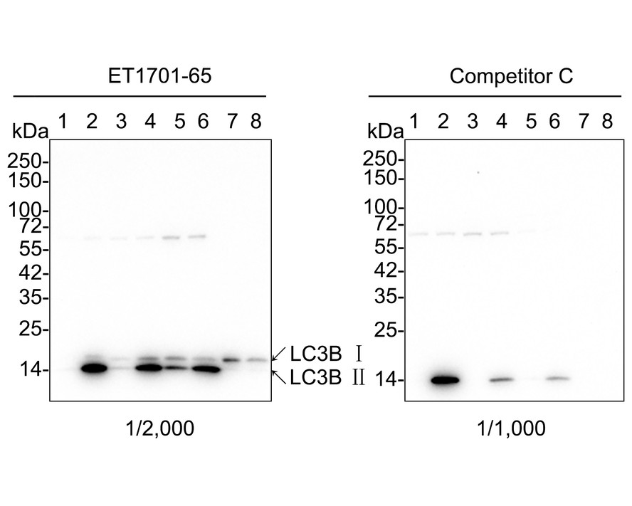
Western blot analysis of LC3B on different lysates with Rabbit anti-LC3B antibody (ET1701-65) at 1/2;000 dilution and competitor's antibody at 1/1;000 dilution. Lane 1: HeLa cell lysate Lane 2: HeLa treated with 50μM Chloroquine for 18 hours cell lysate Lane 3: C2C12 cell lysate Lane 4: C2C12 treated with 50μM Chloroquine for 18 hours cell lysate Lane 5: C6 cell lysate Lane 6: C6 treated with 50μM Chloroquine for 18 hours cell lysate Lysates/proteins at 20 µg/Lane. Predicted band size: 14/16 kDa Observed band size: 14/16 kDa Exposure time: 3 minutes; 4-20% SDS-PAGE gel. Proteins were transferred to a PVDF membrane and blocked with 5% NFDM/TBST for 1 hour at room temperature. The primary antibody (ET1701-65) at 1/2;000 dilution and competitor's antibody at 1/1;000 dilution were used in 5% NFDM/TBST at 4℃ overnight. Goat Anti-Rabbit IgG - HRP Secondary Antibody (HA1001) at 1/50;000 dilution was used for 1 hour at room temperature.
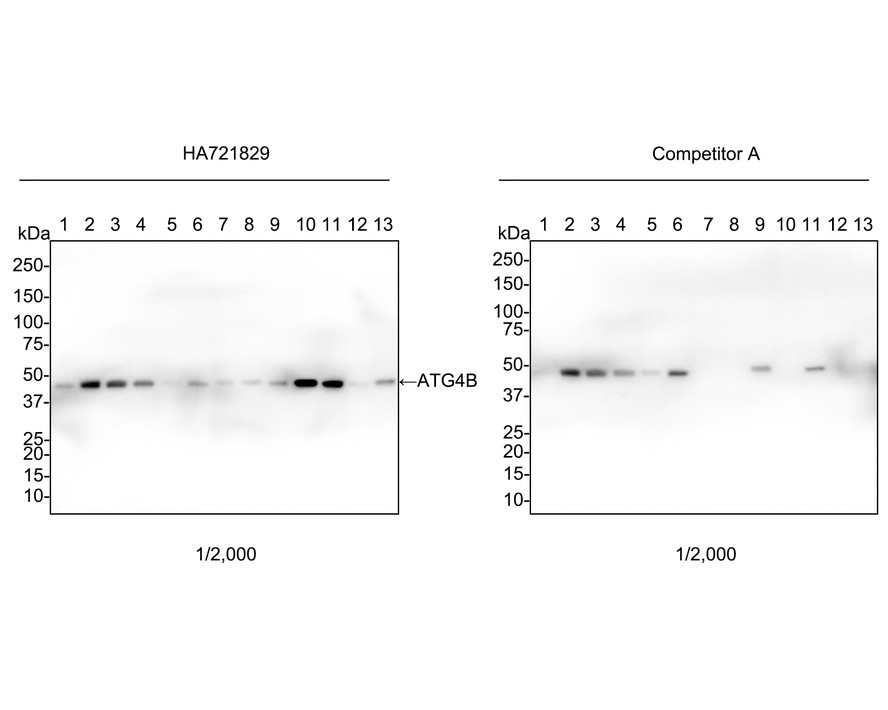
Western blot analysis of ATG4B on different lysates with Rabbit anti-ATG4B antibody (HA721829) at 1/2;000 dilution and competitor's antibody at 1/2;000 dilution. Lane 1: HeLa cell lysate Lane 2: Jurkat cell lysate Lane 3: 293T cell lysate Lane 4: MCF7 cell lysate Lane 5: K-562 cell lysate Lane 6: COS-1 cell lysate Lane 7: NIH/3T3 cell lysate Lane 8: C2C12 cell lysate Lane 9: PC-12 cell lysate Lane 10: Mouse brain tissue lysate Lane 11: Rat brain tissue lysate Lane 12: Mouse kidney tissue lysate Lane 13: Mouse colon tissue lysate Lysates/proteins at 30 µg/Lane. Predicted band size: 44 kDa Observed band size: 47 kDa Exposure time: 42 seconds; 4-20% SDS-PAGE gel. Proteins were transferred to a PVDF membrane and blocked with 5% NFDM/TBST for 1 hour at room temperature. The primary antibody (HA721829) at 1/2;000 dilution and competitor's antibody at 1/2;000 dilution were used in 5% NFDM/TBST at 4℃ overnight. Goat Anti-Rabbit IgG - HRP Secondary Antibody (HA1001) at 1/50;000 dilution was used for 1 hour at room temperature.
Western blot analysis of Apg7 on different lysates with Rabbit anti-Apg7 antibody (ET1610-53) at 1/2;000 dilution. Lane 1: HeLa cell lysate Lane 2: Jurkat cell lysate Lane 3: THP-1 cell lysate Lysates/proteins at 15 µg/Lane. Predicted band size: 78 kDa Observed band size: 70 kDa Exposure time: 3 minutes; 4-20% SDS-PAGE gel. Proteins were transferred to a PVDF membrane and blocked with 5% NFDM/TBST for 1 hour at room temperature. The primary antibody (ET1610-53) at 1/2;000 dilution was used in 5% NFDM/TBST at 4℃ overnight. Goat Anti-Rabbit IgG - HRP Secondary Antibody (HA1001) at 1/50;000 dilution was used for 1 hour at room temperature.
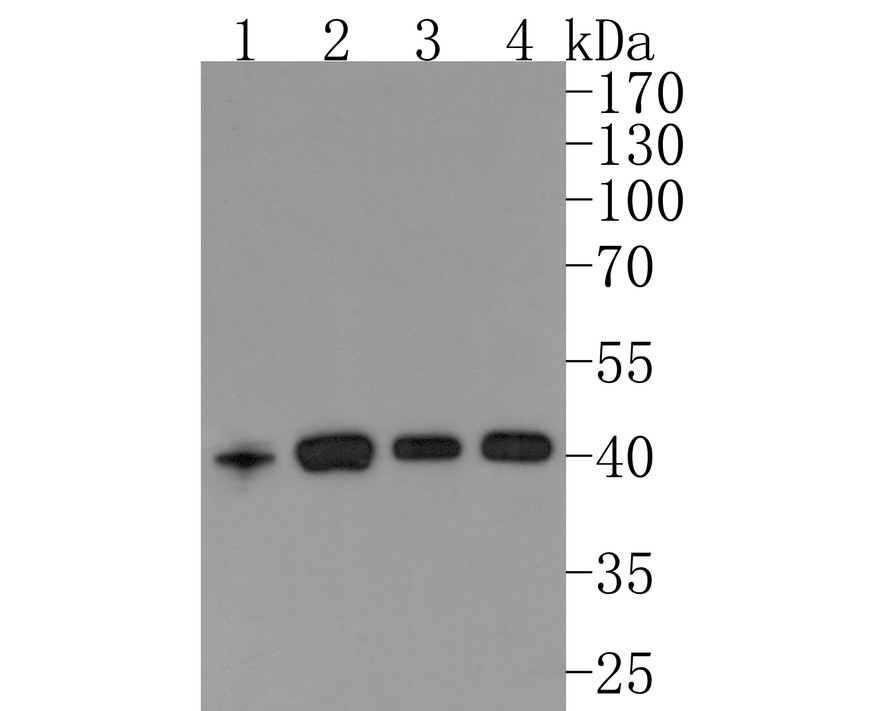
Western blot analysis of ATG4A on different lysates with Rabbit anti-ATG4A antibody (HA721010) at 1/500 dilution. Lane 1: K562 cell lysate; 10 µg/Lane Lane 2: Jurkat cell lysate; 10 µg/Lane Lane 3: Mouse kidney tissue lysate; 20 µg/Lane Lane 4: Rat kidney tissue lysate; 20 µg/Lane Predicted band size: 45 kDa Observed band size: 41 kDa Exposure time: 2 minutes; 10% SDS-PAGE gel. Proteins were transferred to a PVDF membrane and blocked with 5% NFDM/TBST for 1 hour at room temperature. The primary antibody (HA721010) at 1/500 dilution was used in 5% NFDM/TBST at room temperature for 2 hours. Goat Anti-Rabbit IgG - HRP Secondary Antibody (HA1001) at 1:200;000 dilution was used for 1 hour at room temperature.